
Ultrasound examinations Warsaw
At MDT Medical, we offer state-of-the-art ultrasound examinations for adults and children - from abdominal ultrasound, diagnostics of the urinary system and reproductive organs, to thyroid or breast ultrasound. Thanks to our experienced team and advanced equipment, we ensure that abnormalities of the internal organs can be diagnosed quickly and precisely, without a referral and without unnecessary waiting.
4.5/5.0

5.0/5.0

9.5/10

What are the ultrasound examinations like at MDT Medical?
01
Professionalism and experience
Ultrasound examinations at MDT Medical are performed by doctors with many years of experience in the diagnosis of children and adults - with a focus on patient comfort and safety.
02
Modern equipment
We use high-end ultrasound equipment to accurately assess internal organs, tissues and the circulatory system - even in the youngest patients.
03
Individual approach to the patient
We tailor each examination to the patient's age, condition and needs - unhurriedly, with explanation of each stage and results.
04
Quick access to test results
The results of the ultrasound examination are discussed immediately afterwards. If necessary, we immediately refer for further diagnosis or consultation.
Discover the full range of ultrasound examinations at MDT in Warsaw
The examination can detect even small amounts of fluid in the pleural cavities, which is crucial in the diagnosis of inflammation, circulatory failure or complications after trauma. Ultrasonography allows a quick and safe assessment of the lungs and pleura without radiation exposure.
Ultrasound examination with assessment of blood flow in the renal arteries. Helps detect vascular narrowing that can lead to vascular hypertension or renal dysfunction. Indicated, among other things, when hypertension is difficult to control or vascular lesions are suspected.
Study of blood flow in the arteries supplying the intestines - when gastrointestinal ischaemia is suspected.
Helpful in the diagnosis of vasculitis (e.g. giant cell arteritis) - especially in people 50+ with frequent headaches.
Non-invasive brain examination in infants - performed through the open fontanel, it is used to assess development and possible abnormalities.
An extended diagnostic package including three different ultrasound examinations in one visit. An excellent choice for those needing a comprehensive health assessment - such as abdominal, urinary and prostate. No need for multiple appointments.
A comprehensive diagnostic package that allows two different ultrasound examinations to be performed during one visit. This is a convenient option for patients who want to check more than one organ - such as the abdomen and thyroid. Short duration, fast results and greater diagnostic comfort.
Assessment of wrist structures - helpful in the diagnosis of injuries, cysts, degenerative and inflammatory changes.
Assessment of wrist structures - helpful in the diagnosis of injuries, cysts, degenerative and inflammatory changes.
Salivary gland ultrasound is a rapid, safe and non-invasive imaging study that assesses the size, structure and presence of lesions in the parotid, submandibular and sublingual salivary glands.
Ultrasound of both ankle joints allows diagnosis of injuries, sprains, chronic pain, swelling, inflammation and overload changes in these joints.
Hip imaging test - used for pain, injury, degenerative changes.
Hip joint imaging study - used for pain, injury, degenerative changes.
Preventive hip examination in infants - allows early detection of dysplasia.
Ultrasound of the ankle joint allows diagnosis of injuries, sprains, chronic pain, swelling, inflammation and overload changes in this joint.
Ulnar joint ultrasound is a quick, safe and non-invasive imaging study performed to diagnose injury, pain, swelling, inflammation and strain changes in the elbows.
Ulnar joint ultrasound is a quick, safe and non-invasive imaging study performed to diagnose injury, pain, swelling, inflammation and strain changes in the elbow.
Ultrasound of both knee joints is a quick, safe and non-invasive imaging study that assesses the soft tissue structures within the knees - including tendons, ligaments, synovial membranes, synovial bursae, meniscus and the presence of joint fluid.
Ultrasound of the knee joint is a quick, safe and non-invasive imaging study that assesses the soft tissue structures within the knee - including tendons, ligaments, synovial membrane, synovial bursae, meniscus and the presence of fluid in the joint.
Shoulder joint ultrasound is a quick, safe and non-invasive imaging study that assesses the structure of the soft tissues of the shoulders - including the rotator cone tendons, the subacromial bursa, the tendon of the long head of the biceps and the subacromial space.
An ultrasound of the shoulder joint is a quick, safe and non-invasive imaging study that assesses the structure of the soft tissues of the shoulder - including the rotator cone tendons, the subacromial bursa, the tendon of the long head of the biceps and the subacromial space.
Pelvic ultrasound is a quick, safe and non-invasive imaging study that assesses the organs located in the lower abdominal cavity - in women mainly the uterus and ovaries, and in men the prostate gland and urinary bladder.
Chest ultrasound is a quick, safe and non-invasive imaging study that assesses the structures within the chest - primarily the pleura, pleural space, chest wall and the presence of fluid or inflammatory changes. This examination is particularly useful in the diagnosis of dyspnoea, chronic cough, chest pain and complications from infections.
Prostate ultrasound is a rapid, non-invasive and safe imaging study that assesses the size, shape and structure of the prostate gland. It is an essential diagnostic tool in the evaluation of prostatic hyperplasia (BPH), inflammation, focal lesions and urinary disorders.
Assessment of blood flow and detection of aneurysms in the abdominal aorta and iliac arteries.
Examination of the uterus and ovaries performed through the abdominal shell - used, for example, in patients unsuitable for vaginal ultrasound.
Doppler ultrasound of the hepatic arteries is a non-invasive, safe and very precise imaging study that assesses blood flow in the vessels supplying the liver.
Doppler ultrasound of the carotid arteries is a non-invasive, quick and safe test that assesses blood flow in the arteries that supply blood to the brain - mainly the carotid and vertebral arteries.
Doppler ultrasound of the arteries of the upper limbs is a non-invasive, safe and painless examination to assess the condition of the arterial vessels in the hands.
Examination of blood flow in the veins of the legs - helpful, among other things, when thrombosis or varicose veins are suspected.
Blood flow test in the veins of both legs - helpful for suspected thrombosis or varicose veins, among other things.
Assessment of patency and function of veins in the hand - used, for example, for oedema, thrombosis or vascular problems.
Assessment of patency and function of hand veins - used, for example, for oedema, thrombosis or vascular problems.
Doppler ultrasound of the arteries of the upper limb is a non-invasive, safe and painless examination to assess the condition of the arterial vessels in the hand.
Doppler ultrasound of the lower limb arteries is a non-invasive, safe and painless examination to assess the condition of the arterial vessels in the legs.
Doppler ultrasound of the arteries of the lower limb is a non-invasive, safe and painless examination to assess the condition of the arterial vessels in the leg.
Abdominal aortic Doppler ultrasound is a non-invasive, safe and painless imaging study that assesses the diameter of the aorta and the blood flow within it.
Ultrasound of the popliteal fossa is a quick, safe and non-invasive examination that assesses structures at the back of the knee, such as blood vessels, tendons, ligaments and the presence of cysts, inflammation or thrombosis.
Ultrasound of lymph nodes is a non-invasive, safe and quick imaging study that assesses the size, shape and structure of lymph nodes located in the neck, groin, axilla or abdominal cavity, among others.
Urinary ultrasound is a basic, non-invasive imaging study that is used to diagnose diseases of the urinary system. It allows assessment of the kidneys, bladder and prostate gland (prostate) in men.
Soft tissue ultrasound is a non-invasive, rapid and safe diagnostic method to assess the condition of muscles, tendons, ligaments, subcutaneous tissue and lymph nodes.
Thyroid ultrasound is a basic, non-invasive imaging study used in the diagnosis of thyroid disease. It allows the size, shape, structure and echogenicity of the thyroid gland to be accurately assessed and the presence of focal changes such as nodules, cysts or inflammatory infiltrates to be detected.
Neck ultrasound is the primary examination used in the diagnosis of structures located in the neck. It allows an accurate assessment of the lymph nodes, thyroid gland, salivary glands and soft tissues.
Testicular ultrasound is a basic, non-invasive imaging test used in the diagnosis of diseases of the testes, epididymis and spermatic cord. With ultrasound, it is possible to detect cancerous lesions, inflammation, cysts and other pathologies at an early stage that can affect a man's health and fertility.
Abdominal ultrasound is a safe, non-invasive and rapid diagnostic method to assess the internal organs located within the abdominal cavity. The examination allows precise assessment of the liver, gallbladder, pancreas, spleen, kidneys, urinary bladder and large blood vessels.
Doppler ultrasound is a non-invasive ultrasound examination to assess blood flow in blood vessels. It is an essential tool in the diagnosis of diseases of the venous and arterial systems, such as venous insufficiency, thrombosis or atherosclerosis.
Breast ultrasound is a non-invasive, safe and painless imaging study that assesses the structure of the breast gland and surrounding tissues.
Want to take charge of your health? Take the first step - make an appointment at MDT Medical
We cooperate with insurance companies







What do our patients say?

Over 3,200+ positive reviews about our facility


Dr Barbara Matuszkowiak
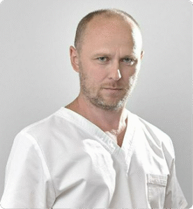
Dr Rafał Wasiak

Stanisław Zwięgliński

Maria Kościuszko, MD, PhD

Izabela Knyziak-Mędrzycka, MD, PhD
Dr Rafał Wasiak
See what issues you can report to us about.
Abdominal pains
Recurrent or sudden abdominal pain can indicate problems with the digestive tract, liver, pancreas or bile ducts.
Suspected kidney or gallbladder stones
Ultrasound can detect deposits in the urinary tract or gallbladder - a common cause of colic or pain on the right side of the abdomen.
Urinary disorders
Urinary retention, frequent urination or soreness may indicate urinary tract disease - ultrasound will assess the kidneys, bladder and prostate gland.
Enlargement of the abdominal circumference
Excessive abdominal enlargement may indicate the presence of fluid in the peritoneal cavity, tumours or cirrhosis.
Abnormal laboratory test results
Increased levels of liver enzymes, urea or creatinine often need to be supplemented by diagnostic imaging.
Yellowing of the skin and eyes
This symptom may indicate liver, biliary tract or gastrointestinal disease - abdominal ultrasound is the first-choice examination.
Testicular pain or swelling
In case of sudden pain, enlargement or asymmetry of the scrotum - ultrasound helps to exclude testicular torsion, hydrocele or tumours.
What should the preparation for the ultrasound examination look like?
An ultrasound examination is safe, painless and non-invasive, but in order for it to be efficient and provide reliable results, it is worth preparing for it. We recommend that you arrive on time for the examination, wearing loose clothing that makes it easier to expose the area of the body being examined. If you have previous imaging or laboratory results - please bring them with you.
Depending on the type of ultrasound (e.g. abdominal, bladder, reproductive organs), the registration staff will give you specific recommendations - e.g. the need to be fasting or to fill your bladder. If you have any concerns, please contact our registration - we will be happy to provide detailed information.
Frequently asked questions
What is ultrasound and what is ultrasound?
Ultrasound, or ultrasonography, is an imaging test that uses high-frequency sound waves. These waves penetrate the patient's body and bounce off the internal organs, creating their image on the screen of the ultrasound machine. This examination is completely safe - it does not use radiation, so it can even be performed on pregnant women and children.
What does the ultrasound examination look like - is it painful?
The ultrasound examination is painless and usually lasts from a few to several minutes. The doctor applies a special gel to the patient's skin, which improves the conductivity of the ultrasound waves, and then moves the camera head over the examined area. The result of the examination is available immediately and can be discussed on site.
What are the indications for an ultrasound examination?
Indications for ultrasound include a wide range of symptoms and conditions - including abdominal pain, suspected diseases of the kidneys, gallbladder, liver, pancreas, thyroid or reproductive system. Ultrasound is also often used for follow-up after trauma and in the diagnosis of abnormal laboratory test results.
What to eat before an abdominal ultrasound and what to avoid?
No food should be consumed 6 hours before the abdominal ultrasound examination. It is also advisable to avoid foods that cause bloating, such as fizzy drinks, dark bread, legumes or raw vegetables. On the day of the examination, you should not smoke or chew gum. It is also advisable to limit your intake of coffee and strong tea.
What is an acoustic shadow on ultrasound and is it a cause for concern?
An acoustic shadow is the phenomenon of an area of shadowing behind a structure that does not transmit ultrasound waves - this could be, for example, a stone in the gallbladder or kidneys. The presence of an acoustic shadow does not necessarily mean anything dangerous, but always requires interpretation by a doctor who will assess its significance in the context of the patient's symptoms and other examination findings.